New zooming technique reveals cell electric circuit for first time
31 October 2017

Cell biologists have used a new super-resolution microscopy technique to be able to observe molecular-level reactions for the first time.
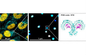
In a paper published in Science Signalling, a team from the University of Reading and Goethe University Frankfurt used a new super-resolution optical microscopy technique to see how Toll-Like Receptors (TLRs), which act like molecular electric circuits to control the flow of signals to a cell. The team have been looking at how a process, called dimerization, decides between life and death of cells and regulates the immune response.
The level of detail achieved by super-microscopy is still not sufficient to make single receptor molecules in a tiny protein dimer visible, so the researchers developed a sophisticated analysis method to improve the optical signal. In this way they were able to ‘zoom in’ on the super-resolution images and examine if TLR4 was present as a monomer or a dimer. The researchers could also detect if chemical signals from different pathogens modulated the receptors’ patterns.
"This process has only ever been observed indirectly, and to see the images that the new microscopic technique provides is very exciting" - Dr Darius Widera, University of Reading
100 times more zoom
Dr Darius Widera, a Cell Biologist from the University of Reading said:
“Using this exciting new technique we were able to observe a particular Toll-like Receptor, TLR4, carry out dimerization on a molecular level for the first time. This process has only ever been observed indirectly, and to see the images that the new microscopic technique provides is very exciting. The process has allowed us to zoom in on TLRs at more than 100 times the power of a standard microscope to provide incredibly detailed information of what the process begins looks like.”
Dr Graeme Cottrell, a biochemist from the University of Reading said: We know that these TLRs are able to police a cell’s fate through forming a dimer molecule. By switching on and off like a light switch, they send signals that can fight harmful bacteria, viruses and the like when they interact. In this new study, we observed how the presence of a chemical signal from several pathogens led to different response from TLR4, which we would have not been able to do without the new super-resolution technique.”
Treating cancer tumour cells
All cells in the human body communicate by sending and receiving chemical signals. The recognition of these signals is achieved by specialised proteins on the cell surface referred to as receptors. These receptors act as molecular switches that convey signals from the surface into biological responses which can be as diverse as cell survival or cell death.
Previous studies carried out by the consortium composed of the research groups led by Dr Widera, Dr Cottrell (both University of Reading, School of Pharmacy) and Prof Heilemann (Goethe University Frankfurt, Germany) have also showed that activation of the TLR4 by different chemical signals can either induce proliferation of brain cancer cells or lead to controlled death of these tumour cells.
Professor Mike Heilemann from the Institute of Physical and Theoretical Chemistry at Goethe University Frankfurt said:
“It is conceivable that this approach will help us in future to understand better the fundamental biological processes that regulate the immune system in health and disease. At the same time, this microscopy method is also applicable to other membrane proteins and many similar questions.”
Full citation: Carmen L. Krüger, Marie-Theres Zeuner, Graeme S. Cottrell, Darius Widera, Mike Heilemann: Quantitative single-molecule imaging of TLR4 reveals ligand-specific receptor dimerization, Science Signaling, doi: 10.1126/scisignal.aan1308
